Polarized light microscopy reveals what ordinary brightfield illumination cannot: the optical properties of crystals, minerals, fibers and other ordered materials. It is a core technique in geology and the material sciences, and a standard tool in education, research and industrial laboratories.
This article explains how polarized light microscopy works, what it shows, where it is used, and what to look for in a polarizing microscope.
What is polarized light microscopy?
Polarized light microscopy is a contrast technique that uses polarized light to study how a specimen interacts with light. A polarizer below the specimen lets through light vibrating in a single direction. An analyzer above the specimen is set at 90 degrees to the polarizer, so with no specimen in place the field of view is dark. This setting is called crossed polarizers.
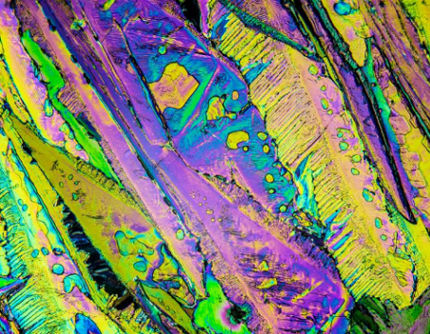
Isotropic materials, such as glass or minerals with a cubic crystal structure, have the same refractive index in every direction and stay dark between crossed polarizers. Anisotropic, or birefringent, materials split the light into two rays that travel at different speeds. When the rays recombine at the analyzer they interfere, and the specimen appears bright and often brightly colored against the dark background.
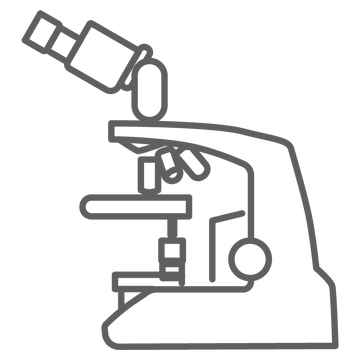
Polarizing microscopes are also called petrographic or geological microscopes, after their best known application.
How a polarizing microscope works
A polarizing microscope adds a set of dedicated components to a transmitted light microscope:
- Polarizer and analyzer: the polarizer sits below the condenser and the analyzer in the light path above the objectives. Both can be rotated or set to the crossed position.
- Rotating stage: a circular stage graduated in degrees turns the specimen between the polarizers, so extinction positions and angles can be observed and measured.
- Strain-free optics: objectives and condensers made to be free of internal stress. Ordinary optics can add birefringence of their own, which would show up as false contrast.
- Centerable objectives: each objective is centered to the axis of the rotating stage, so the specimen stays in the field of view as the stage turns.
- Compensators: a full wave (lambda) plate, a quarter wave plate and a quartz wedge slide into the light path to measure retardation and to identify the slow and fast directions in a crystal.
- Bertrand lens: brings the back focal plane of the objective into view for conoscopic observation, where interference figures help determine whether a crystal is uniaxial or biaxial and its optic sign.
What makes polarized microscopy stand out?
Polarized light turns optical properties into visible contrast. Interference colors relate to the thickness of the specimen and the strength of its birefringence. Extinction angles, pleochroism, relief and interference figures each add information about the identity and orientation of a crystal.
This makes polarized light microscopy both qualitative and quantitative. A trained observer can identify a mineral, reveal stress in a molded part, or measure retardation with a compensator, all on the same instrument and usually without staining the specimen.
Where polarized light microscopy is used
- Geology and petrography: identification of minerals in rock thin sections and study of rock texture.
- Materials science: examination of polymers, fibers, ceramics and crystalline materials, and of stress patterns in transparent parts.
- Chemistry and crystallography: observation of crystal habit, birefringence and crystal growth.
- Building materials: study of concrete, aggregates and other construction materials.
- Biology: contrast in naturally ordered structures such as starch grains, plant fibers and other birefringent biological material.
- Education: teaching optical mineralogy and crystal optics in earth science and materials courses.
What to look for in a polarizing microscope
- Strain-free objectives and condenser, so the only birefringence you see belongs to the specimen.
- A graduated 360 degree rotating stage and objectives that can be centered to it.
- A rotating polarizer and an analyzer that can be adjusted and removed from the light path.
- Compensator slots with full wave, quarter wave and quartz wedge plates.
- A Bertrand lens for conoscopic work.
- Even LED illumination with Köhler alignment and intensity control.
- A crosshair eyepiece for alignment and angle measurements.
- A trinocular head if you need to document what you see with a camera.
LABOMED polarized microscopes
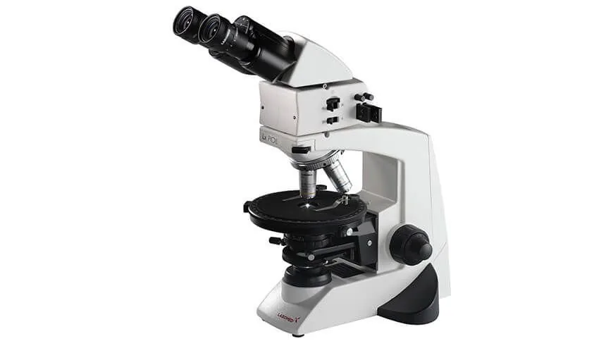
The LABOMED® Lx POL is a polarizing microscope built for these requirements. Its SF series strain-free, infinity corrected DIN plan achromatic objectives (4x, 10x and 40x, with an optional 100x oil objective) sit in a reverse angle quadruple nosepiece, and each objective can be centered individually.
- Magnification from 40x to 400x with the standard objectives, and 1000x with the optional 100x oil objective.
- A 160 mm stage that rotates through 360 degrees in one degree increments.
- A rotating polarizer and an adjustable analyzer with centration screws, a focusable Bertrand lens, and lambda, quarter lambda and quartz wedge plates.
- A 0.9 NA centerable, strain-free condenser.
- LED illumination with intensity control and a Köhler field diaphragm.
- Siedentopf binocular or trinocular head, 30 degree inclined, with focusable 10x/20 mm eyepieces and a crosshair in one eyepiece.
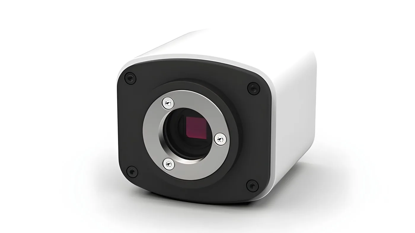
- On the trinocular head, C-mount adapters for the LABOMED Vega, Vega 4K and iVu Touch cameras.
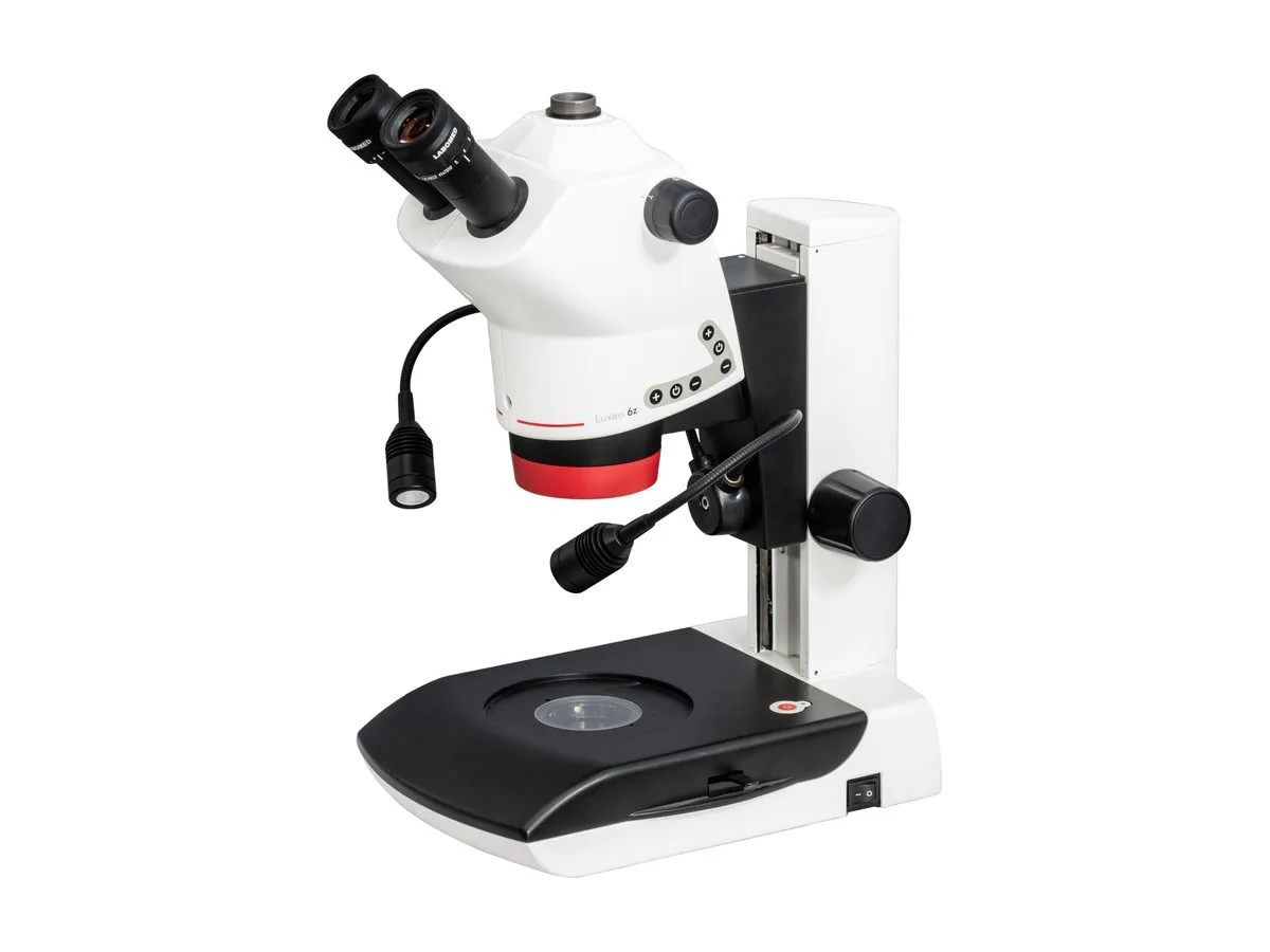
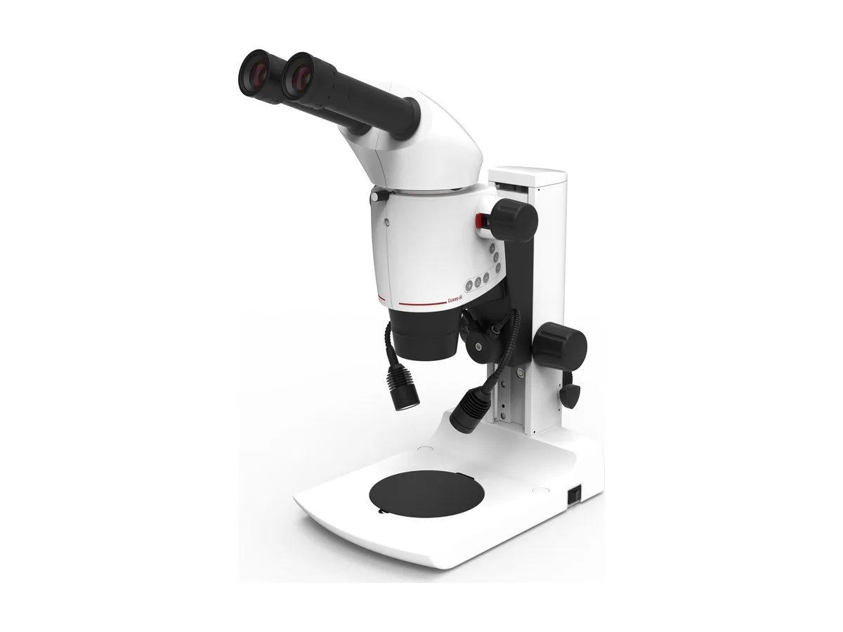
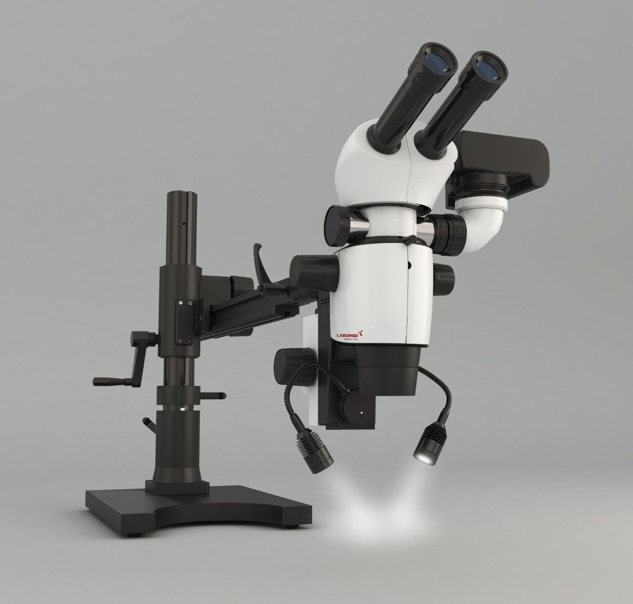
For low magnification work on larger samples, the Luxeo 6Z and Luxeo 6i stereo microscopes accept an optional polarizing attachment.
LABOMED microscopes are manufactured by Labotech and sold and distributed by Labo America, which can help you configure the Lx POL for your laboratory or classroom.
FAQs
What is polarized light microscopy used for?
It is used to identify and study birefringent materials: minerals in rock thin sections, crystals, polymers, fibers, ceramics and building materials. It is also used to reveal stress in transparent materials and to teach optical mineralogy.
What is the difference between a polarizing microscope and a standard microscope?
A polarizing microscope adds a polarizer, an analyzer, a rotating graduated stage, strain-free optics, compensator slots and usually a Bertrand lens. A standard brightfield microscope has none of these, so it cannot show birefringence reliably.
Why do polarizing microscopes need strain-free objectives?
Stress inside ordinary glass makes the optics slightly birefringent. Between crossed polarizers this shows up as false contrast that can be mistaken for the specimen. Strain-free optics keep the background dark, so only the specimen produces contrast.
What does a Bertrand lens do?
A Bertrand lens lets you observe the back focal plane of the objective. This conoscopic view shows interference figures, which help determine whether a crystal is uniaxial or biaxial and what its optic sign is.
 APPLICATION · ELECTRONICSHow Inspection Microscopes Are Used in Electronics and PCB Assembly Quality Control?
 BUYING GUIDE · INDUSTRIALChoosing the Right Microscope for Industrial and Material Sciences
 PRODUCT · STEREOLuxeo 6i: The Shining Beacon in Industrial & Medical Stereo Microscopy