ACROSS DENTISTRY
Microscopes across dentistry.
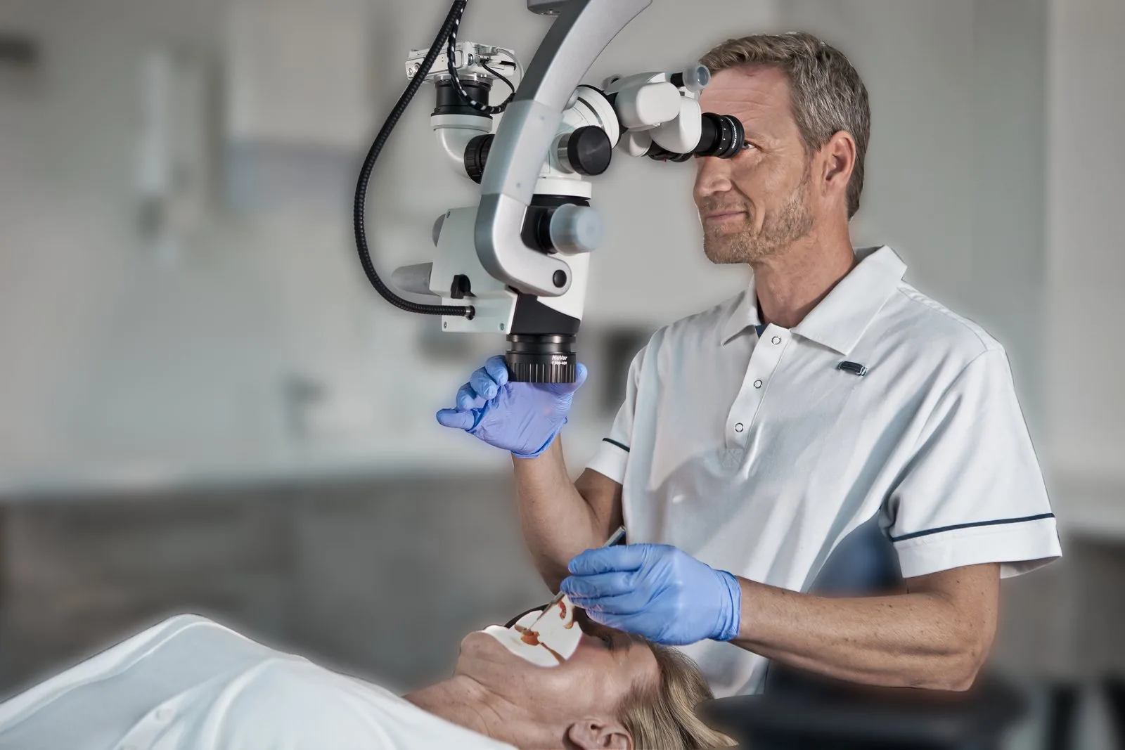
The operating microscope earned its place in endodontics. Today it serves the whole practice, from the first examination to the final restoration, and from the operatory to the teaching lab. Here is what each discipline asks of it, and how LABOMED® dental microscopes answer.
Endodontics
Canal anatomy sits below the reach of unaided vision. Intense coaxial light and high magnification help clinicians locate calcified canals, remove obturation materials and find cracks, and microsurgical root-end technique has reported markedly higher success than the traditional approach.
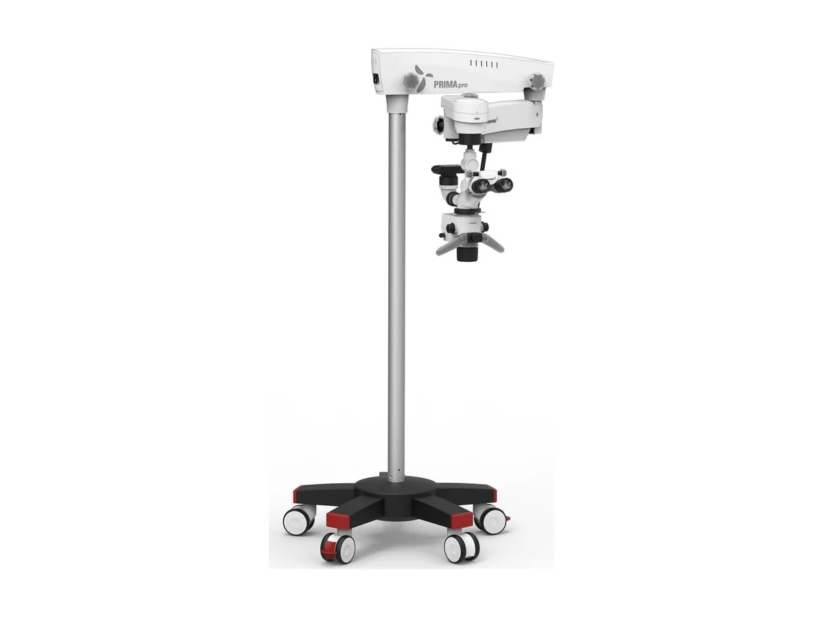
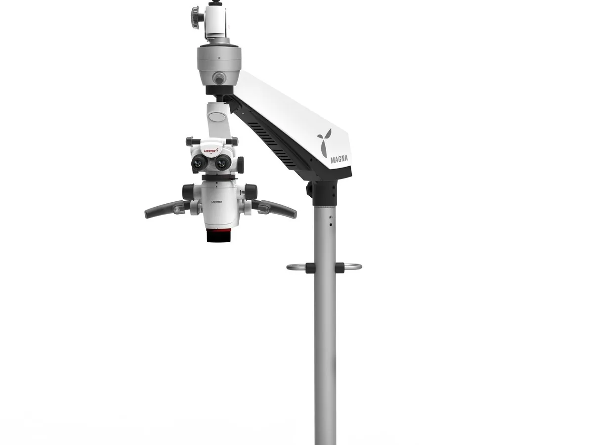
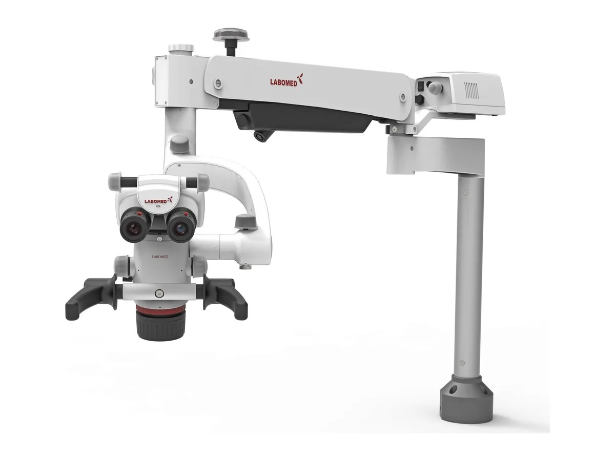
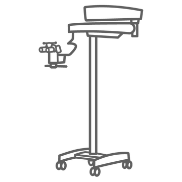
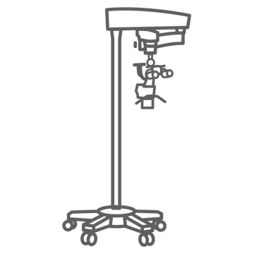
HOW LABOMED ANSWERSPrima Pro pairs a six-step apochromatic magnification changer with 50 W LED light rated at 200,000 lux at 200 mm. Magna adds a continuous 4x to 25x zoom with a selectable click-stop, so magnification follows the procedure rather than fixed steps.
Restorative and cosmetic dentistry
Restorative work is decided at the margin. Magnification brings precision to removing old restorations and evaluating marginal gaps, and in a laboratory study crown preparations refined under the microscope showed tighter margins than those refined under loupes.
HOW LABOMED ANSWERSYellow and green filters are built in: yellow for working with light-cured resin, green for blood-free contrast. Prima Pro adds TrueLux, a warmer color temperature for clinicians who prefer the look of halogen light.
Prosthodontics
Fixed prosthodontics is judged at the finish line. Under the microscope, small differences in marginal ridge height and the seating of a restoration become visible, and in a study of veneer preparation the microscope gave prosthodontists better posture than loupes or the unaided eye.
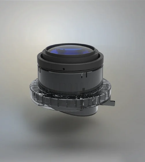
HOW LABOMED ANSWERSNuVar variable working distance objectives let you refocus by turning the objective instead of moving the microscope: NuVar 10, f 300 to 400 mm, is standard on Prima Pro and Magna, with NuVar 20, f 200 to 400 mm, as the option.
Periodontics
Periodontal plastic surgery rewards gentle handling of thin tissue and exact closure. A meta-analysis comparing microsurgical and conventional technique for gingival recession found better root coverage, esthetics and recovery with microsurgery, and a 2024 systematic review traces the microscope through grafting and ridge preservation.
HOW LABOMED ANSWERSThe green filter heightens soft tissue contrast. On Prima Pro and Magna, an electromagnetically balanced arm holds the head where you leave it, without drift, through long grafting procedures.
Oral surgery and implantology
Surgical fields in the mouth are small and deep. Clinicians use the microscope to inspect extraction sockets for residual root fragments or an oro-antral communication, and microsurgical principles now extend to implant placement, soft tissue management and bone augmentation.
HOW LABOMED ANSWERSNuVar 20 gives Prima Pro and Magna working distances from f 200 to 400 mm, leaving room for instruments and assistance. An optional assistant viewer bridge on Prima Pro lets the assistant follow the field.
Pediatric dentistry
Primary teeth are small and appointments are short. Pediatric dentists in a 2021 survey agreed that the microscope helps locate canals in primary teeth and that a live image can support behavior management, while noting that adoption is still early and that some children may find the equipment unsettling.
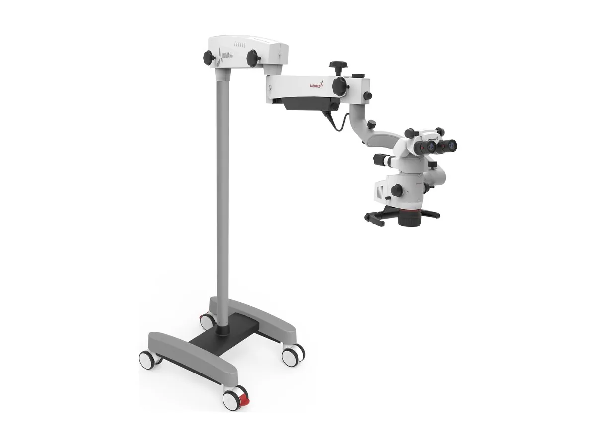
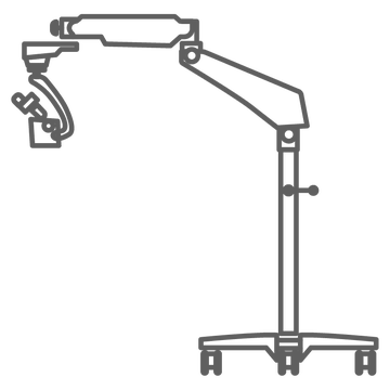
HOW LABOMED ANSWERSPrima Lite carries its 27 W LED inside the microscope body, with no fiber optic cable, on a compact, fluid arm with a park mode. The Smartphone Adapter streams the view wirelessly, so a parent can see what the clinician sees.
Hygiene and diagnostics
Many problems show themselves late: an enamel crack, early caries, deposits left on a root surface. Magnification and focused light make it easier to identify cracks, inspect caries and the tissue around it, and see residual irritants after debridement.
HOW LABOMED ANSWERSPrima Lite brings more than 100,000 lux of LED light and a five-step apochromatic changer, 0.4x to 2.5x, to a compact stand. Magna's continuous zoom lets you survey at low power and close in on a finding without stepping through fixed settings.
Patient communication and documentation
A finding the patient can see is easier to explain than one they must take on trust. Camera integration captures high-resolution images for records and patient conversations, and the photographic record also supports communication with referring dentists.
HOW LABOMED ANSWERSEvery LABOMED® dental microscope accepts beam splitters with C-mount, DSLR and Smartphone Adapter options, and the Atlas+ 4K camera. Prima Pro also offers a rear-facing beam splitter.
Ergonomics for the clinician
Dentistry is practiced in long, static postures, and near vision declines with age. In one study, dentists from age 40 showed significantly lower near acuity, and acuity was better with the microscope than with loupes at comparable magnification; in another, the microscope improved head, neck and elbow posture more than loupes.
HOW LABOMED ANSWERSA 0 to 210° inclinable binocular head lets you sit upright while the optics adapt to the patient. Prima Pro has two-button controls for left- and right-handed users, and Magna has a brake release button on each handle.
Dental education
Microscope skills take practice, and the profession expects them: microscope use is taught in every CODA-accredited advanced education program in endodontics, and reviewers urge dental schools to bring magnification into the core curriculum.
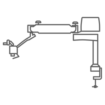
HOW LABOMED ANSWERSPrima Trainer is a table-mounted surgical training microscope with five-step apochromatic optics, LED illumination and a 0 to 210° head, used in dental, ENT and ophthalmology training labs. Camera adapters let an instructor show the class what a student sees.