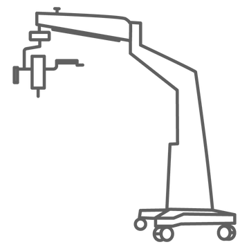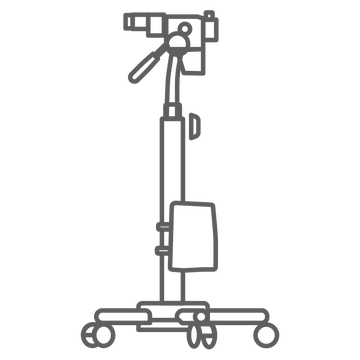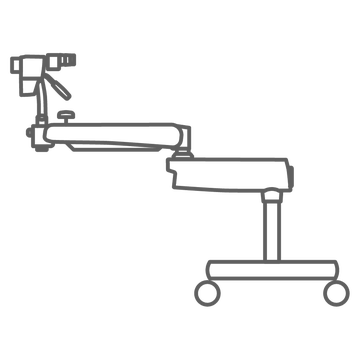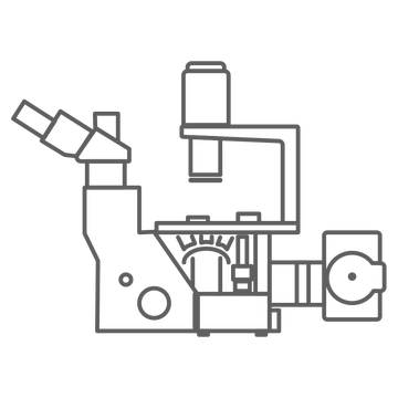
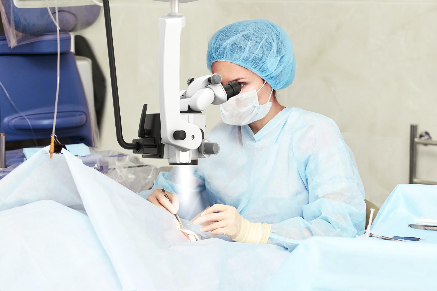
Understanding how surgical microscopes can be used in stereotaxic examinations helps researchers set up procedures where small errors in position change the result. In a stereotaxic procedure, a target inside the brain is reached using three-dimensional coordinates measured from landmarks on the skull. A surgical microscope gives the operator the magnified, well lit and stable view needed to find those landmarks and to work precisely at them.
This article explains the role of the microscope in stereotaxic surgery in mice and rat models, what to look for in a microscope for a stereotaxic frame, and which LABOMED® systems suit stereotaxic research benches.
The role of surgical microscopes in stereotaxic procedures
Stereotaxic surgery places an injection needle, cannula, electrode or optical fiber at a defined position in the brain. The animal is held in a stereotaxic frame, the skull is leveled, and coordinates are taken from skull landmarks, most commonly bregma and lambda.
Every step depends on what the operator can see. A surgical microscope supports this work with:
- Magnification to identify sutures and landmarks on a small skull
- Binocular stereo viewing for depth perception when lowering instruments
- A working distance long enough to clear the frame, manipulator arms and drill
- Even, shadow-free LED illumination of the exposed skull
- A stable mount that holds the view still during long procedures
Stereotaxic surgery in mice
The mouse skull is small, so landmarks such as bregma and lambda, the sutures and the surface vessels are hard to read without magnification. A surgical microscope helps the operator:
- Identify bregma and lambda and take accurate reference coordinates
- Check that the skull is level before measuring
- Place burr holes precisely and avoid surface vessels
- Watch the needle or cannula as it is lowered and confirm its position
Stereotaxic surgery rat models
Rat models offer larger anatomical targets, but procedures can be longer and may involve several implants. Here the microscope matters as much for comfort and consistency as for magnification:
- A comfortable, upright viewing position reduces operator fatigue in long sessions
- Lower magnification and a wide field of view help orientation across the whole skull
- Higher magnification supports fine work at the burr hole and around implants
- A long working distance leaves room for implants, anchor screws and cement
Stereotaxic surgery mouse protocol and visualization accuracy
A typical stereotaxic surgery mouse protocol includes these steps, each of which benefits from a clear magnified view:
- Animal positioning in the frame and leveling of the skull
- Skull exposure and cleaning
- Identification of bregma and lambda
- Burr hole drilling at the calculated coordinates
- Injection or implantation
- Closure and recovery
Your institution's approved protocol defines the procedure itself. The microscope helps every operator carry out the same steps in the same way.
Optical advantages that support stereotaxic accuracy
Magnification and resolution
A magnification changer lets the operator start at low magnification to orient on the skull, then step up to identify landmarks and work at the burr hole. Apochromatic, coated optics keep the image sharp and color true across the range.
Illumination control
Coaxial LED illumination lights the field along the viewing axis, so there are no shadows from instruments or the frame. Adjustable intensity helps avoid glare on a wet skull surface.
Ergonomics and stability
Binocular viewing on a tilting head keeps the operator upright. A well balanced arm or boom keeps the optical axis steady and lets the head be moved out of the way between steps.
Using a Microscope with a Stereotaxic Frame
Using a microscope with a stereotaxic frame comes down to space and stability. Check these points before you choose a system:
- Working distance: enough room between the objective and the skull for drills, manipulator arms and injection systems.
- Reach and positioning: an arm or boom that places the head over the frame without moving the animal, and parks it clear when not in use.
- Mounting: a table clamp or a heavy base that keeps vibration away from the frame.
- Field of view: wide enough to see bregma and lambda together at low magnification.
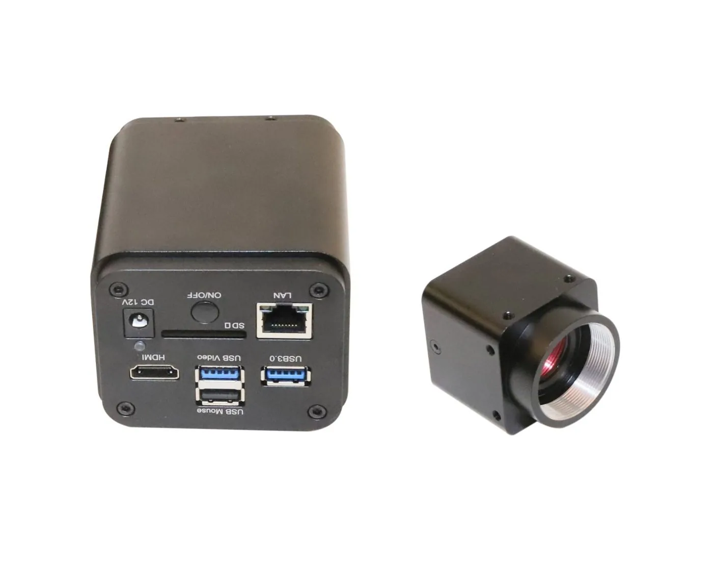
- Documentation: a camera port or beam splitter to record procedures for training and lab records.
Training, reproducibility, and research quality
Microscopes also help laboratories train new researchers and keep procedures consistent between operators.
- A shared magnified view of the same landmarks
- Images and video recorded through a camera for protocol records and teaching
- Consistent illumination and magnification settings from one session to the next
Limitations and considerations when selecting a surgical microscope
Key factors to weigh include:
- Bench space and the footprint of the frame
- Working distance against the magnification you need
- Whether the lab needs a camera now or later
- Whether the microscope will also be used for other procedures or for training
LABOMED microscopes for stereotaxic research
LABOMED offers two practical routes for a stereotaxic bench.
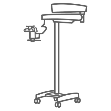
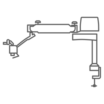
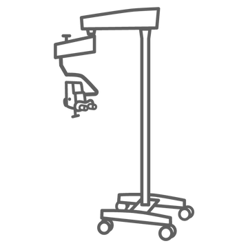
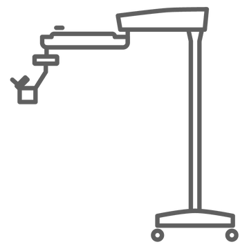
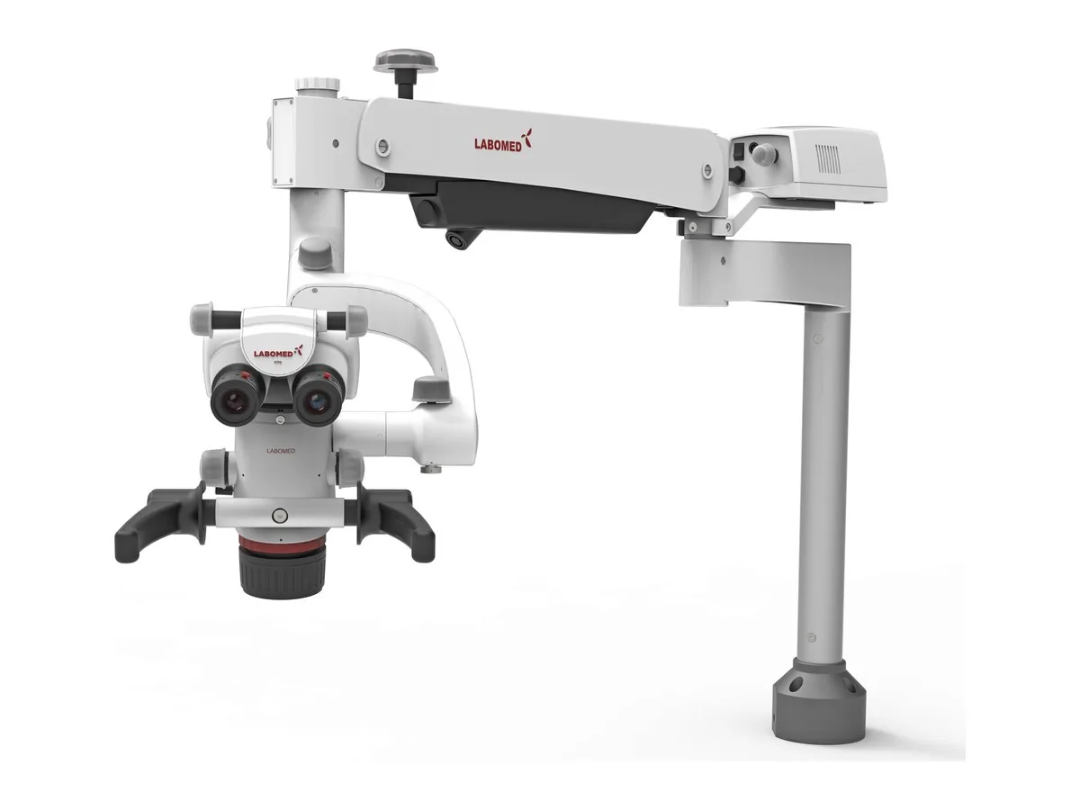
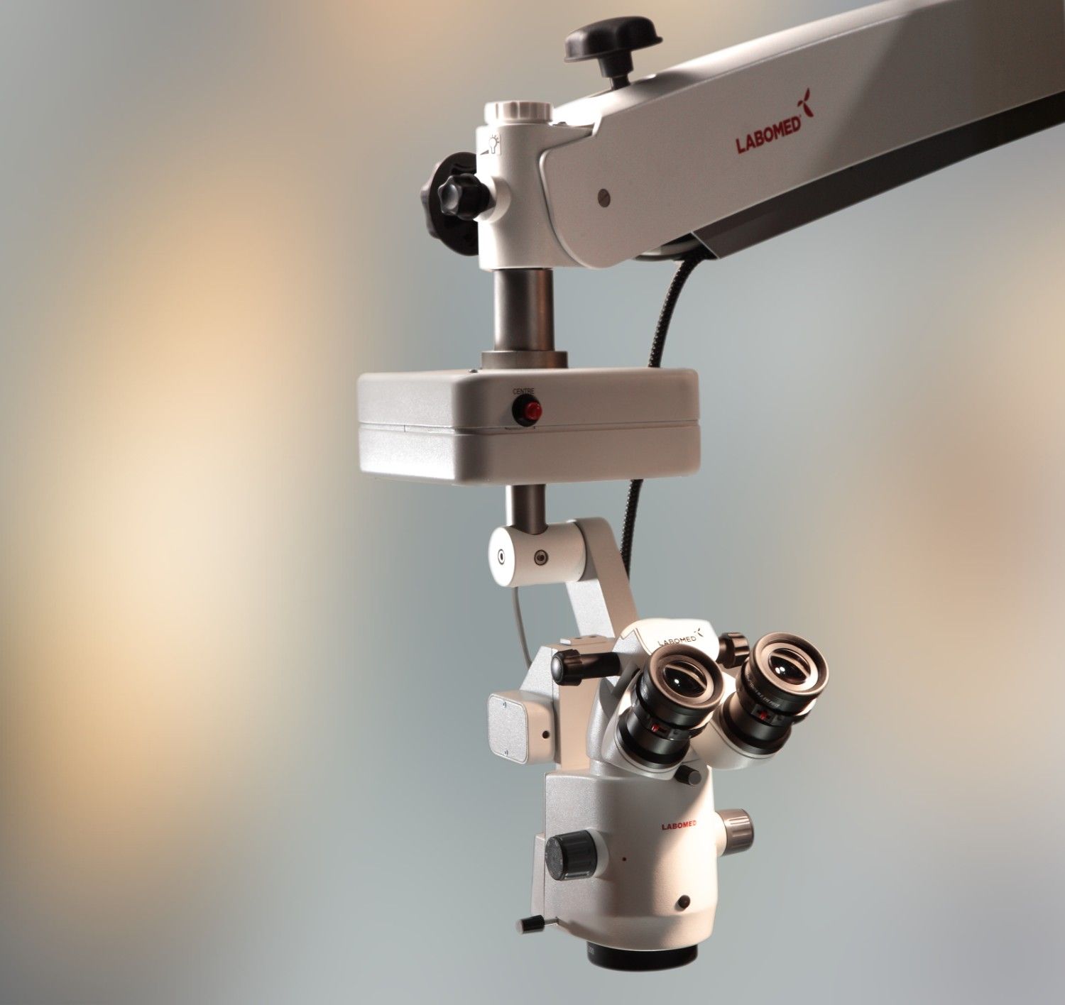
- Prima Trainer: a surgical training microscope on a table mount with a C-clamp, with a 120 degree carrier and 550 mm of arm travel. It has a 5 step apochromatic magnification changer (0.4x, 0.6x, 1.0x, 1.6x and 2.5x) with MaxLite coated optics, a 0 to 210 degree tilting head, and a 50 W LED that delivers 125,000 lux. The standard f 250 mm objective is focusable, with f 200, 300 and 400 mm objectives as options, and a beam splitter accepts C-mount cameras including the Atlas+ 4K.
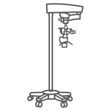
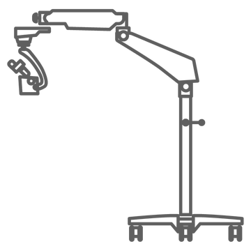
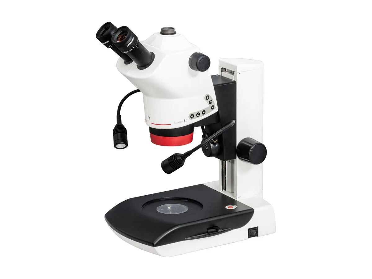
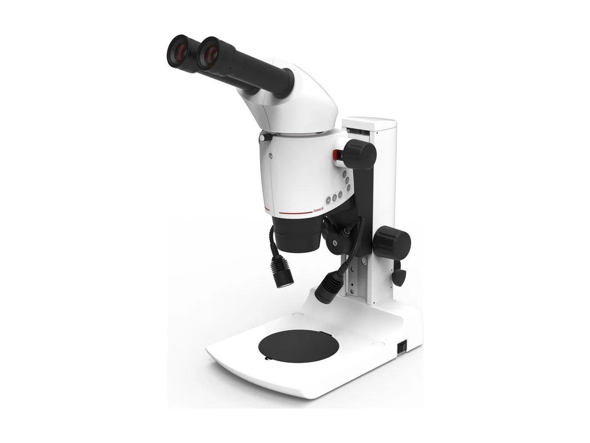
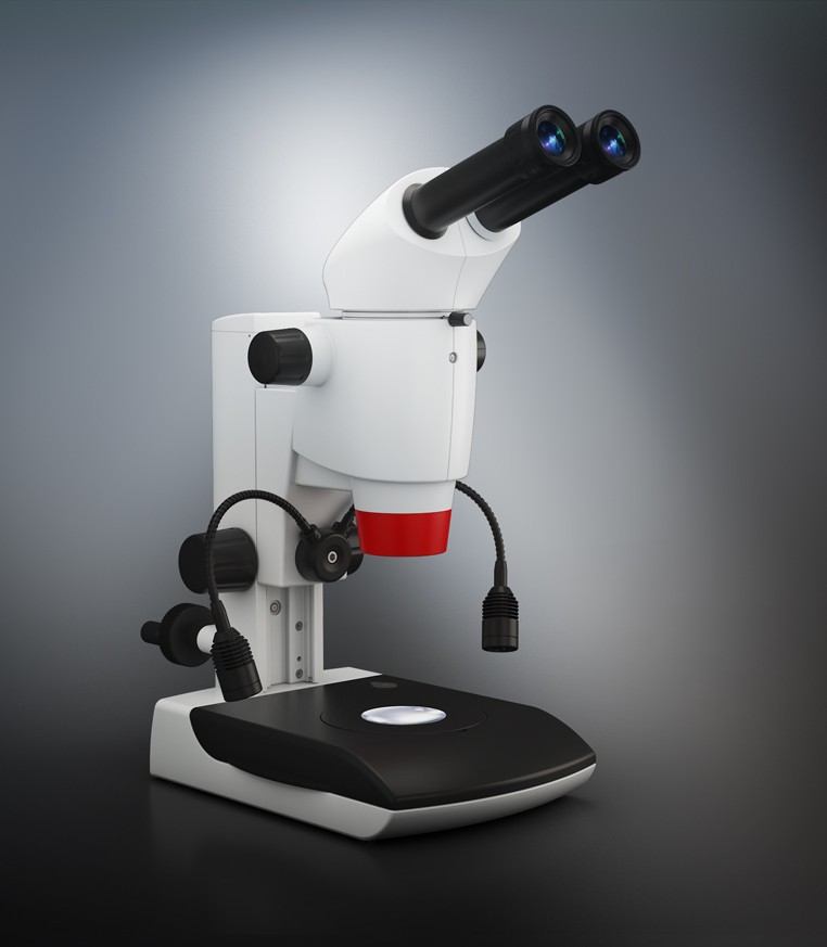
- Luxeo 6Z and Luxeo 6i: stereo zoom microscopes with 8x to 50x continuous magnification and a 110 mm working distance, available on boom stands. The Luxeo 6Z combines an LED ring light with dual gooseneck lights; the Luxeo 6i adds coaxial LED illumination, a heavy duty boom with crank adjustment and an optional 0 to 210 degree tilting head.
See the microsurgical training application page for more on skills labs and research benches. LABOMED microscopes are manufactured by Labotech and sold and distributed by Labo America, which can help you match a configuration to your frame and bench.
FAQs
Why use a surgical microscope for stereotaxic surgery?
A surgical microscope magnifies the skull landmarks used to take stereotaxic coordinates, lights the field evenly and gives binocular depth perception, which helps the operator place burr holes, needles and implants precisely and consistently.
What magnification do you need for stereotaxic surgery in mice?
Most work combines low magnification for orientation over the whole skull with higher magnification for landmarks and burr holes. A magnification changer or zoom covers both without changing the setup.
What working distance does a microscope need for a stereotaxic frame?
It needs enough room for the drill, manipulator arms and injection system between the objective and the skull. The Prima Trainer works at f 250 mm as standard, with f 200, 300 and 400 mm objectives as options, and the Luxeo 6Z and Luxeo 6i stereo microscopes work at 110 mm.
Can I record stereotaxic procedures through the microscope?
Yes. A beam splitter on a surgical microscope, or a C-mount adapter on a stereo microscope, lets you attach a camera to record images and video for training and lab records.
Final thoughts: choosing the right solution for your lab
Whether you work with stereotaxic surgery in mice, stereotaxic surgery rat models or other small animal research, the right microscope makes landmarks easier to read and procedures easier to repeat. Choose for working distance and stability first, then magnification range and documentation.

 EXPLAINER · LABCompound Microscope vs Stereo Microscope: Which One Does Your Lab Need?
 APPLICATION · IVFHow Microscopes Are Used in IVF and Embryology Labs?
 APPLICATION · PHARMAHow Surgical Microscopes Benefit the Field of Pharmaceutical Research?