
Key Takeaways:
- A digital microscope captures the magnified image with a camera sensor and shows it on a screen, so it can be recorded, measured and shared.
- Its core components are the optics, the illumination, the image sensor, the camera electronics, the display and the connections to a computer or network.
- Image quality starts with the optics: a sensor can only record the detail the objective resolves.
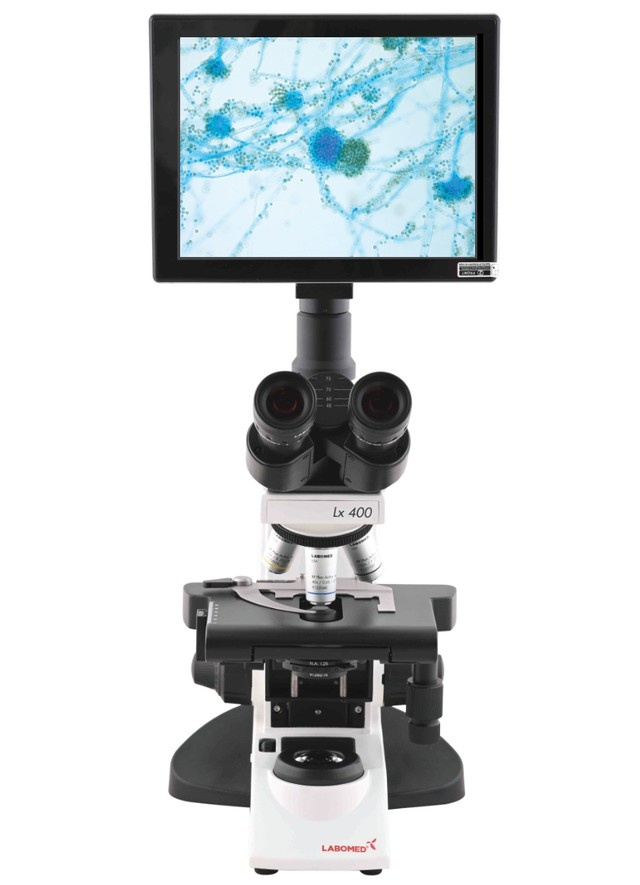
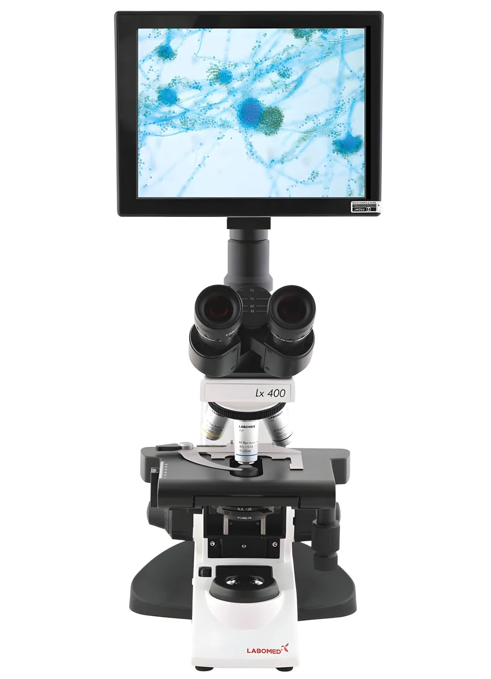
- Many laboratories build a digital microscope by adding a C-mount camera to a trinocular compound or stereo microscope, which keeps the eyepieces for direct viewing.
- LABOMED® offers three C-mount cameras for this: the Atlas+ 4K, the Vega and Vega 4K, and the iVu Touch with its own screen.
A digital microscope sends its image to a camera sensor and a screen, instead of or as well as to the eyepieces. The magnified view becomes a digital image that you can save, measure, annotate and show to a whole room at once.
This guide explains how a digital microscope works, what its core components do, which features matter in daily use, and how digital and optical viewing compare.
What This Blog Covers
- How a digital microscope works and what makes it different
- The key components that make up a digital microscope system
- Modern digital microscope features and how they support real workflows
- How digital and optical microscopes compare in practice
- Where digital microscopes are used today
- How LABOMED approaches digital microscopy
- FAQs
A digital microscope is a system that uses a camera and image sensor to capture magnified images and display them on a monitor, a computer or a built-in screen. Users work with the image digitally, which makes recording, analysis and sharing part of the normal workflow.
How a Digital Microscope Works
The optical path starts the same way as in any microscope. Light from the illuminator passes through or reflects from the specimen, and the objective forms a magnified image.
In a digital system, that image is projected onto an image sensor instead of only into the eyepieces. The sensor converts light into an electronic signal, the camera electronics turn it into a color image, and the result appears on a screen as a live view.
From there, the image can be frozen, captured as a still, recorded as video, measured with software or sent to another display. In a classroom, the whole group follows the same field of view. In a laboratory or on an inspection bench, each observation can be stored with the sample record.
Looking at the Components That Make It Work
The components of a digital microscope are straightforward. What matters is how well they are matched to each other.
- Optics: the objective and the zoom or magnification system set resolution, magnification and working distance.
- Illumination: LED light gives even, stable lighting with consistent color, which matters as much to a camera as to the eye.
- Image sensor: a CMOS sensor converts the optical image into pixels. Its size and pixel count set how much of the field is captured and how much detail the file holds.
- Camera electronics: exposure, white balance and noise reduction are handled in the camera, which outputs a live image.
- Display: a monitor or television over HDMI, a computer screen over USB, or a screen built into the camera itself.
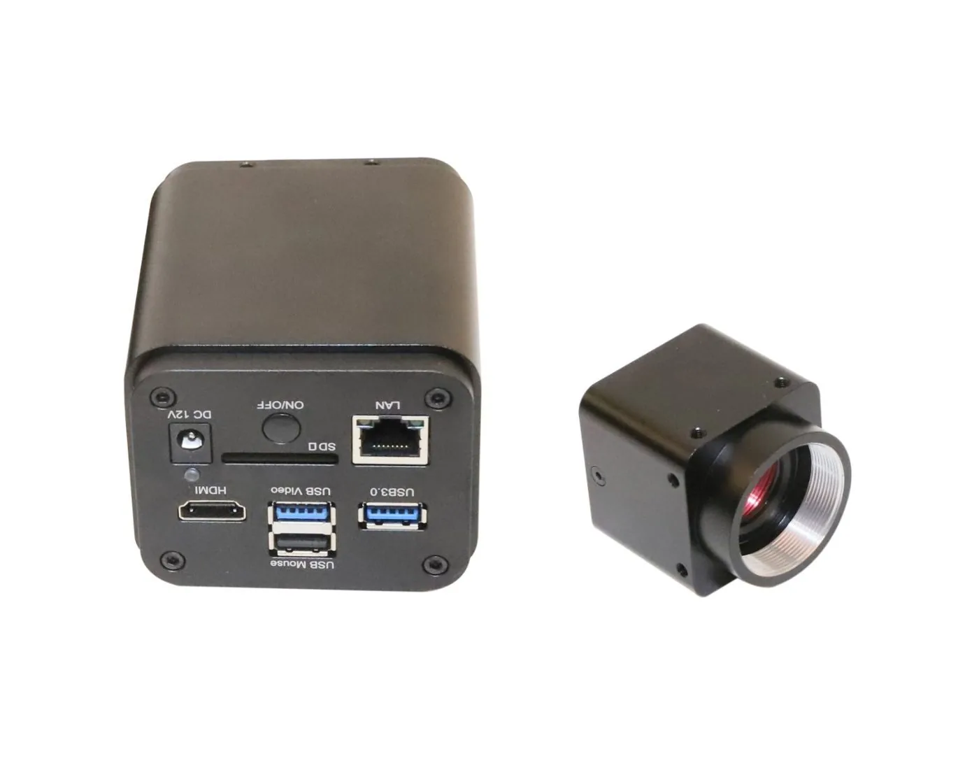
- Connectivity and storage: USB, HDMI, network connections and SD card storage move images into reports, records and teaching material.
- Camera adapter: on a modular system, a C-mount adapter couples the camera to the microscope and matches the image to the sensor.
What Defines Modern Digital Microscope Features
The features that matter most are the ones that change daily work.
- Resolution and frame rate: a 4K sensor records fine detail for documentation, and a high frame rate keeps the live image smooth while the specimen moves or focus changes.
- Standalone operation: cameras with their own controls and HDMI output run without a computer, which suits classrooms and production benches.
- Measurement and annotation: software tools add scale, dimensions and notes to images.
- Color reproduction: consistent white balance and color matter in clinical, materials and dental documentation.
- Sharing: network or Wi-Fi output sends the live image to other screens and other people.
This is what makes digital imaging valuable wherever documentation matters as much as observation.
Digital vs Optical Microscopes
An optical microscope delivers the image straight to the eye through the eyepieces. The view is immediate, a stereo microscope gives natural depth perception, and nothing stands between the user and the specimen.
A digital microscope adds a recorded, shareable image. Several people can watch at once, results can be documented and reviewed later, and measurements are stored with the image.
The two are not mutually exclusive. A trinocular microscope with a camera keeps direct viewing through the eyepieces and adds a digital image on the third port, so users can work through the eyepieces and record at the same time.
Where Digital Microscopes Fit Today
- Education: instructors show the live image on a classroom screen so every student sees the same field, and capture images for lessons and assessments.
- Clinical and research laboratories: images document findings, support second opinions and build a repeatable record.
- Inspection and materials: stored images give quality control a traceable record of each part, joint or surface.
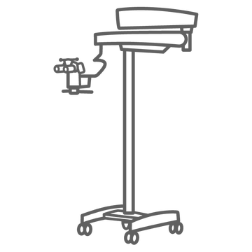
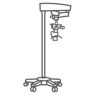
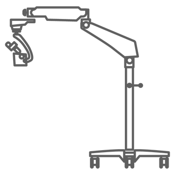
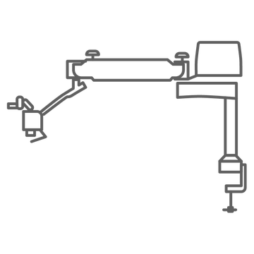
- Dental and surgical microscopy: a camera on the microscope records procedures for patient communication, case records and training.
LABOMED and Digital Microscopy
LABOMED® takes a modular approach. You choose the microscope for the work, then add the camera that fits how you need to capture and share images. All three LABOMED cameras connect through a standard C-mount.
- Atlas+ 4K: a compact 4K camera that records video at 30 fps in 3840 x 2160, runs its own operating system with mouse control on a 4K screen, and connects over USB 3.0, HDMI, LAN and SD card.
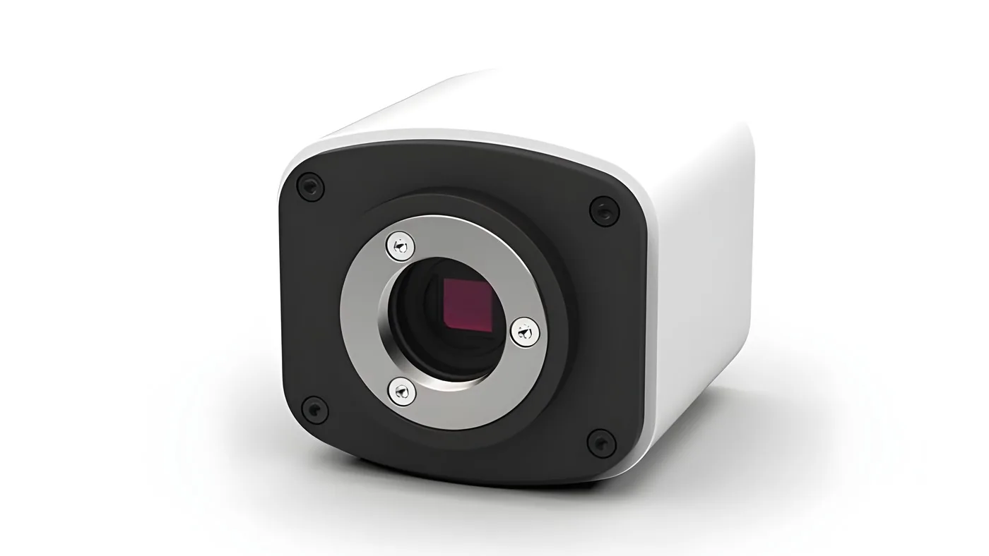
- Vega and Vega 4K: the Vega is a 6 MP camera with HDMI streaming at 30 fps in 1080p, SD card capture, and measurement and settings controlled with a USB mouse. The Vega 4K delivers 3840 x 2160 in real time at 30 fps and adds Wi-Fi.
- iVu Touch: a 5 MP camera built into a screen of nearly 10 inches, with measurement and parameter settings on screen, SD card storage and USB mouse control. It is a complete digital station with no separate monitor or computer.
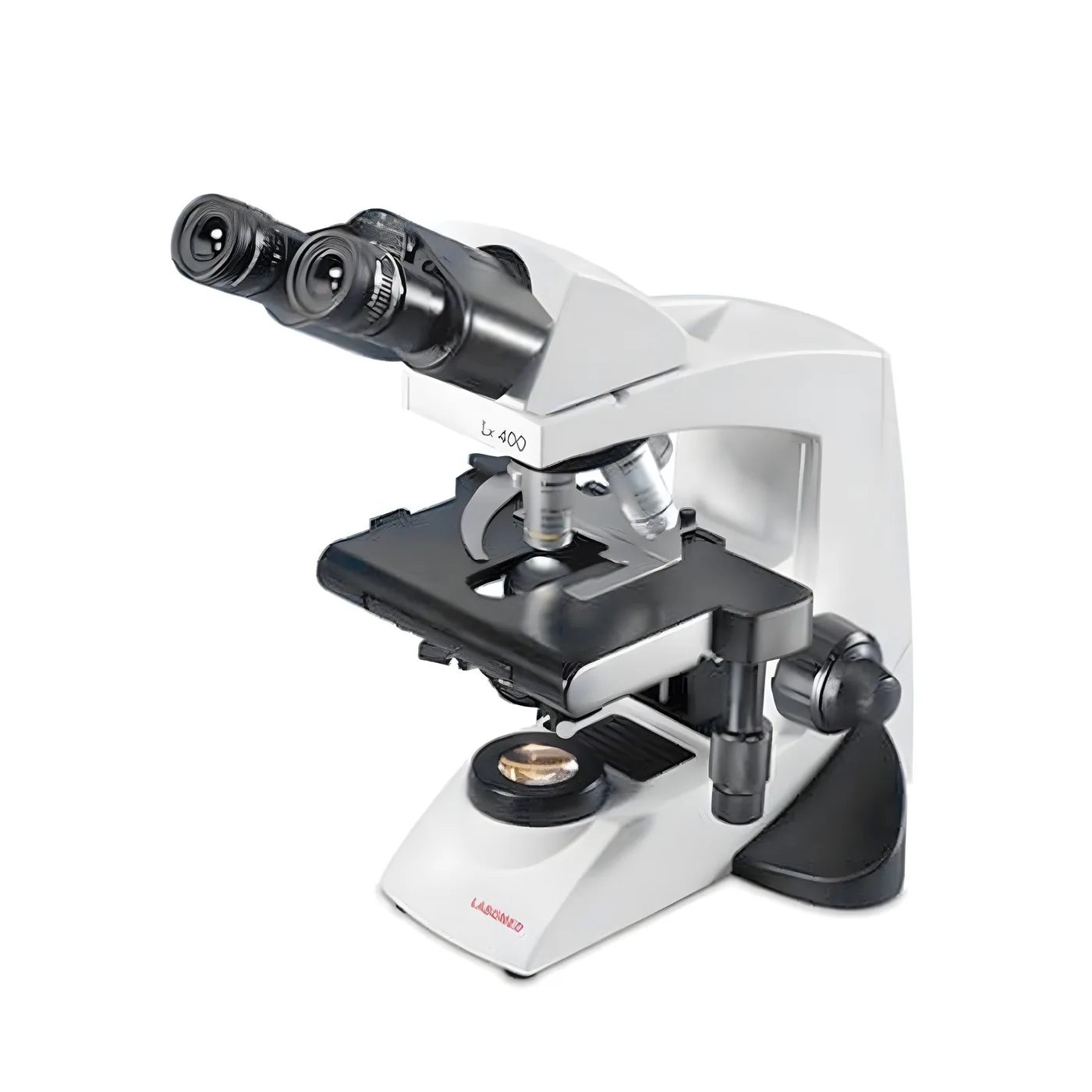
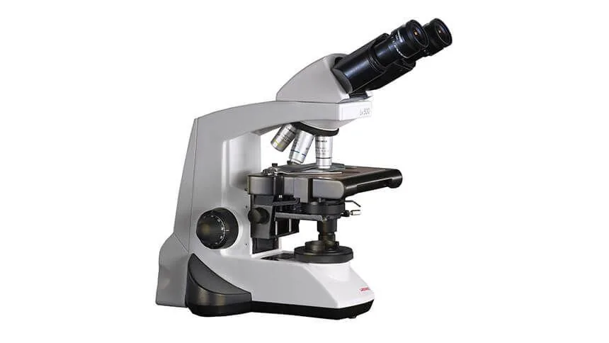
These cameras fit LABOMED compound microscopes with a trinocular head, such as the Lx 400 and Lx 500, through C-mount adapters. LABOMED dental and surgical microscopes take a camera through a beam splitter. Labo America confirms the right adapter for each configuration.
LABOMED microscopes and cameras are manufactured by Labotech and sold and distributed by Labo America.
FAQs
What is a digital microscope used for?
A digital microscope is used wherever you need to see fine detail and also record it. In classrooms, everyone can look at the same image at once. In laboratories and industrial settings, it documents what you see so it can be reviewed, measured or shared later.
What are the components of a digital microscope?
Most systems combine optics, an LED light source, an image sensor, camera electronics and a display, with USB, HDMI or network connections for storage and sharing. On a modular system, a C-mount adapter connects the camera to the microscope.
How does a digital microscope work?
The objective forms a magnified image, which is projected onto an image sensor. The camera turns the signal into a color image and shows it live on a screen, where you can capture stills, record video and take measurements.
What are the benefits of using a digital microscope?
The main benefits are documentation and sharing. Images can be saved with the sample record, measured, annotated and shown to a group, which makes teaching, collaboration and quality records easier.
What is the difference between a digital microscope and an optical microscope?
An optical microscope shows the image only through the eyepieces. A digital microscope also puts the image on a screen, so it can be recorded, analyzed and shared. A trinocular microscope with a camera gives you both.
Conclusion
A digital microscope is defined less by its magnification than by what you can do with the image. Good optics and even LED illumination produce the detail, the sensor and camera electronics capture it, and the display and connections put it to work.
For most laboratories, classrooms and inspection benches, the practical route is a well matched microscope and camera. Start from the specimen and the workflow, choose the optics, then choose the camera that records and shares the image the way your team works.

 EXPLAINER · LABCompound Microscope vs Stereo Microscope: Which One Does Your Lab Need?
 APPLICATION · IVFHow Microscopes Are Used in IVF and Embryology Labs?
 APPLICATION · PHARMAHow Surgical Microscopes Benefit the Field of Pharmaceutical Research?