How to Use a Microscope for ENT: 10 of the Most Common Uses

Key Takeaways:
1. ENT microscopes provide magnified, illuminated views of small and anatomically complex structures.
2. They are used across otology, neurotology, laryngology, skull base surgery, and selected head and neck procedures.
3. Ear surgery remains one of the most established applications for operating microscopes in ENT.
4. Microscopy and endoscopy are not interchangeable. The choice depends on the anatomy, access and procedure.
5. Magnification, illumination, working distance, ergonomics and documentation capabilities should be considered together when selecting an ENT microscope.
Otolaryngology covers a wide range of procedures, but many of them involve anatomy where a clear view of fine structures is essential. This is particularly evident in ear surgery, cochlear implantation, microlaryngeal procedures, and selected skull base and microsurgical applications. ENT microscopes provide the magnification and illumination needed in these settings, while also supporting examination, surgical training, and documentation.
As a manufacturer of medical microscopes, Labomed develops optical systems for clinical environments where visualization, positioning, and practical workflow all need to work together. This guide looks at the main uses of ENT microscopes and where they fit within otolaryngology practice.
What This Blog Covers
1. What Are ENT Microscopes Used For?
3. How ENT Microscopes Improve Visualization
5. Microscope vs Endoscope in ENT
6. How to Match an ENT Microscope to Clinical Use
What Are ENT Microscopes Used For?
An ENT microscope is used when the procedure requires a magnified and well-illuminated view of small anatomical structures. Common applications include otoscopy, ear surgery, cochlear implantation, microlaryngoscopy, selected sinus and skull base procedures, microvascular work, diagnosis, treatment planning, surgical training and documentation.
What Is an ENT Microscope?
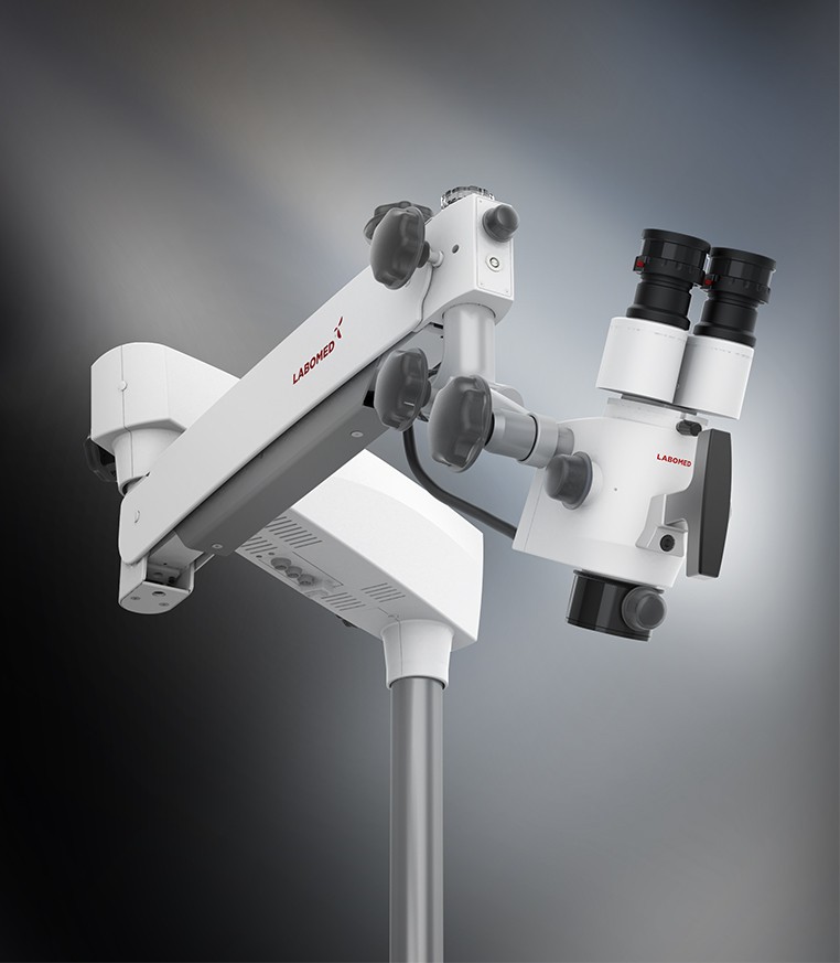
An ENT surgical microscope is designed for procedures where the surgeon needs more than an unaided view of a confined surgical field. The optical system provides magnification while the illumination system directs light into the area being examined or treated.
The value is particularly apparent in otology and neurotology, where structures such as the tympanic membrane, ossicles and middle-ear anatomy occupy a small working field.
Microscopes are also used outside ear surgery. Their role extends into laryngeal procedures, selected head and neck work and microsurgical applications where controlled visualization matters.
How ENT Microscopes Improve Visualization
Magnification is only one part of the equation. A useful operating microscope combines magnification with illumination, optical clarity, depth of field and a working distance suited to the procedure.
For an ENT practice, the relevant question is therefore not simply how much magnification does the microscope provide? It is whether the optical and mechanical configuration suits the procedures being performed.
10 Uses of ENT Microscopes
1. Otoscopy
An operating microscope can provide a more detailed view of the external auditory canal and tympanic membrane than routine unaided examination.
For clinicians already using otoscopy as part of their diagnostic workflow, microscopy can be particularly useful when closer inspection is needed before or during treatment.
2. Ear Surgery
Microscope for ear surgery is one of the most established applications of ENT microscopy.
Procedures involving the middle ear and other delicate structures can require a stable, magnified view of a relatively small operative field. Microscopy is commonly associated with procedures in otology and neurotology where precise visualization is central to the surgical approach.
3. Cochlear Implantation
Cochlear implantation involves work around small structures within the ear, making controlled visualization important throughout the procedure.
An ENT microscope can provide the magnified operative view required during the relevant stages of implantation and associated ear surgery.
4. Laryngoscopy and Microlaryngeal Surgery
Are ENT microscopes used for laryngoscopy? Yes, particularly where direct, magnified visualization is required for microlaryngeal procedures.
Microscopy can be used to examine and treat structures of the larynx, including vocal cord lesions, when the procedure calls for a close operative view.
5. Sinus and Nasal Procedures
Microscopy has applications in selected sinus and nasal procedures, although endoscopy is central to many contemporary sinonasal workflows.
For this reason, the question is less about whether a microscope or endoscope is universally preferable and more about which visualization system provides the access and view required for the specific procedure.
6. Microvascular Surgery
Microvascular procedures demand controlled visualization of very small structures. In selected head and neck and reconstructive procedures, operating microscopy can support the close visual work involved in vessel handling and microsurgical technique.
7. Skull Base Surgery
Skull base procedures can involve narrow anatomical corridors and structures that are difficult to inspect at conventional scale.
Depending on the surgical approach, microscopy may form part of the visualization strategy, particularly when a direct, magnified operative field is required.
8. Head and Neck Procedures
The operating microscope for otolaryngology is not limited to one subspecialty. Selected head and neck procedures can also benefit from magnified visualization when the operative field or anatomy makes fine detail important.
The exact role depends on the procedure and the surgical approach being used.
9. Diagnosis and Treatment Planning
Microscopy can also have a role before surgery. A more detailed view of relevant anatomy can support examination and treatment planning in appropriate clinical situations.
This is particularly relevant when subtle anatomical findings need to be assessed closely rather than viewed only at standard examination scale.
10. Training and Surgical Documentation
ENT microscopes can also become part of the teaching and documentation workflow. With appropriate imaging or video capture, the operative field can be displayed for teaching, recorded for documentation, or reviewed after a procedure.
For practices that train residents or fellows, this can make the microscope useful beyond the surgeon seated at the operating position.
Microscope vs Endoscope in ENT
Microscopy and endoscopy serve different purposes, and many ENT practices use both.
An operating microscope provides a binocular, magnified view through an optical system positioned outside the operative field. This can be particularly useful for procedures involving the ear and other applications where a stable, direct view is required.
An endoscope, by contrast, can be introduced into anatomical spaces and can provide views along angles that may not be accessible through a microscope.
The choice depends on the anatomy, access route, procedure and surgeon's established technique. In some workflows, the two systems are complementary rather than competing technologies.
How to Match an ENT Microscope to Clinical Use
The best ENT microscope for otolaryngology is ultimately the one that fits the procedures and working environment of the practice.
When comparing systems, consider:
1. Magnification levels: The useful range should correspond to the procedures performed regularly.
2. Illumination: Bright, focused illumination is important when working in narrow or deep fields.
3. Working distance: The objective configuration should provide enough space for instruments and comfortable positioning.
4. Ergonomics: The microscope should support a practical operating posture over longer procedures.
5. Imaging: Camera and video options can matter for teaching, documentation and case review.
6. Mounting: Floor, wall or other mounting configurations should suit the available clinical space.
7. Upgrade options: A modular system can provide flexibility as the practice's requirements change.
For Labomed, the focus is on optical performance, illumination and practical configuration across medical microscopy applications. Its microscope portfolio can be explored through the ENT and otolaryngology microscope range.
FAQs
Q. What Are the Main Uses of an ENT Microscope?
A. ENT microscopes are commonly used for ear surgery, otoscopy, cochlear implantation, microlaryngeal procedures, selected sinus and skull base procedures, microvascular work, diagnosis, training, and surgical documentation.
Q. Which ENT Procedures Require an Operating Microscope?
A. The need depends on the procedure and surgical approach. Microscopy is particularly established in ear surgery and is also used in selected laryngeal, skull base, microvascular, and head and neck procedures.
Q. Can an ENT Microscope Be Used for Ear Surgery?
A. Yes. Ear surgery is one of the principal applications of operating microscopes in otolaryngology, particularly where small middle-ear structures require magnified visualization.
Q. Are ENT Microscopes Used for Cochlear Implants?
A. Yes. Microscopy can be used during cochlear implantation and associated otologic procedures where detailed visualization of small anatomical structures is required.
Q. Can ENT Microscopes Be Used for Laryngeal Procedures?
A. Yes. Operating microscopes are used for selected laryngeal and microlaryngeal procedures, including procedures involving vocal cord lesions.
Q. Are ENT Microscopes Used in Sinus Surgery?
A. They can be used in selected sinus and nasal procedures, although endoscopy is widely used in sinonasal surgery. The appropriate system depends on the procedure and surgical approach.
Q. What Is the Difference Between an ENT Microscope and an Endoscope?
A. A microscope provides a magnified optical view from outside the operative field, while an endoscope can be introduced into anatomical spaces to provide views from within them. Both have distinct applications in ENT.
Looking for an ENT microscope for your practice? Explore Labomed's otolaryngology microscopy solutions or contact the team to discuss the configuration suited to your clinical workflow.
Conclusion
The uses of ENT microscopes extend well beyond routine ear examination. From otologic and cochlear procedures to microlaryngeal surgery, skull base work, microvascular applications and surgical training, microscopy remains relevant wherever a close, controlled view of anatomy is required.
For practices evaluating an ENT microscope, the application should drive the specification. Magnification, illumination, working distance, ergonomics, imaging and mounting are most useful when considered in relation to the procedures the microscope will actually support.

© 2010-2026 Labo America, Inc. All Rights Reserved.