How Surgical Microscopes Are Used in Reconstructive and Plastic Surgery?

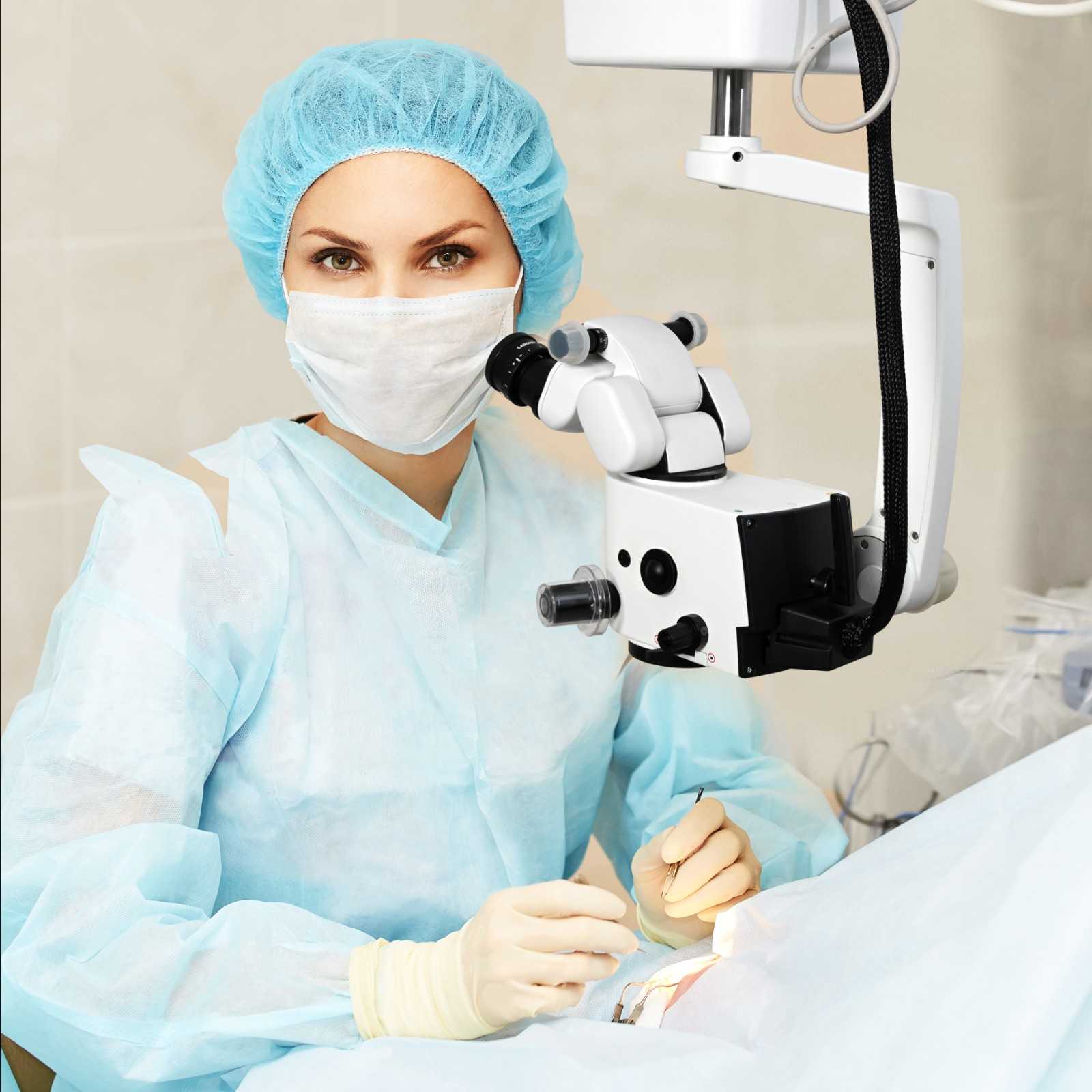
Key Takeaways:
- A surgical microscope for reconstructive and plastic surgery supports visualization during complex microsurgical procedures.
- Free flap reconstruction, microvascular repair, and tissue restoration place unique demands on microscope systems.
- Optical clarity, illumination, depth perception, and ergonomics often matter more than maximum magnification alone.
- Modern operating microscopes for microsurgery are increasingly evaluated for workflow integration, imaging capabilities, and long-term usability.
- The best microscope systems support surgical precision without disrupting established surgical techniques and workflows.
Reconstructive surgery often demands a level of precision that leaves little room for compromise.
Whether performing free flap reconstruction, microvascular tissue transfer, nerve repair, or limb salvage procedures, surgeons frequently move between broader anatomical views and structures measured in millimeters.
In these environments, visualization becomes more than a convenience. It becomes part of the surgical workflow itself.
That is why the surgical microscope for reconstructive and plastic surgery remains a critical component of modern microsurgical practice. The right system supports clarity, consistency, and efficiency throughout complex procedures without becoming a distraction in the operating room.
This guide explores how surgical microscopes are used across reconstructive surgery and what surgeons and healthcare organizations often evaluate when selecting a microscope system.
What This Blog Covers
- How operating microscopes for microsurgery support reconstructive procedures
- The role of visualization in free flap and microvascular surgery
- Key considerations beyond magnification alone
- The importance of ergonomics and workflow integration
- What to evaluate when comparing surgical microscope systems
A surgical microscope for reconstructive and plastic surgery is an advanced visualization system used during microsurgical procedures to provide magnified, illuminated, and stereoscopic views of delicate anatomical structures, supporting precision during tissue reconstruction, vessel repair, and complex surgical procedures.
Why Reconstructive Surgery Places Unique Demands on Visualization
Unlike many surgical specialties, reconstructive microsurgery often requires surgeons to transition between larger anatomical structures and highly detailed microvascular work during the same procedure.
A free flap reconstruction, for example, may involve tissue harvest, flap preparation, vessel dissection, and microvascular anastomosis, each presenting different visualization requirements.
That is why selecting a surgical operating microscope for reconstructive procedures involves more than reviewing technical specifications. The microscope must support multiple stages of surgery while maintaining consistent optical performance throughout.
For many reconstructive teams, the evaluation begins with a practical question:
Can the microscope support the complexity of the procedure without becoming a limitation?
Consistency Matters More Than Maximum Magnification
When evaluating an operating microscope for microsurgery, magnification naturally receives attention.
However, many surgeons place equal importance on how the microscope performs across the entire magnification range.
A microscope should maintain:
- Stable image brightness
- Reliable depth perception
- Consistent focus
- Smooth magnification transitions
- Clear visualization throughout the procedure
The objective is not simply to magnify structures. It is to provide dependable visualization as surgical requirements change from one stage of the procedure to the next.
This becomes especially relevant in reconstructive surgery, where surgeons may move repeatedly between tissue preparation, vessel work, and reconstruction.
Free Flap Procedures Highlight the Importance of Optical Performance
Few applications demonstrate the value of a microsurgery visualization system more clearly than free flap reconstruction.
During microvascular anastomosis, surgeons rely on detailed visualization of small blood vessels and surrounding soft tissue structures. In these situations, optical performance directly influences the user experience.
A high-resolution surgical microscope for anastomosis should provide:
- Sharp vessel visualization
- Consistent illumination
- Reliable stereoscopic imaging
- Natural tissue representation
- Stable depth of field
The goal is to deliver a visual platform that supports the surgeon's preferred technique while maintaining consistency throughout lengthy procedures.
Ergonomics Influence Microsurgical Workflow
Experienced reconstructive surgeons understand that microscope performance extends beyond optics alone.
Positioning flexibility, maneuverability, and viewing comfort all contribute to how effectively a microscope integrates into the operating room.
When evaluating a medical microscope for microvascular surgery, considerations often include:
- Viewing angle flexibility
- Microscope movement and balance
- Access around the surgical field
- Assistant viewing capabilities
- Long-case usability
The most effective systems support the surgical workflow naturally, allowing teams to focus on the procedure rather than the equipment.
The Operating Room Has Changed. So Have Expectations from Microscopes .
Today's reconstructive procedures often involve more than direct visualization.
Recording capabilities, imaging systems, training requirements, and collaborative case review have become increasingly important across hospitals and surgical centers.
As a result, many organizations evaluate microscope systems based on their ability to support:
- Surgical education
- Resident and fellow training
- Procedure documentation
- Case review and collaboration
- Operating room integration
These capabilities increasingly influence purchasing decisions alongside traditional optical considerations.
Supporting Advanced Reconstructive Surgery with Labomed
Labomed Medical Microscopes are designed to support the visualization demands of modern microsurgical and reconstructive procedures.
Built around advanced optical systems, ergonomic design principles, and workflow-focused engineering, Labo America microscope platforms are developed to meet the needs of surgeons performing complex tissue reconstruction, microvascular repair, and soft tissue procedures.
Whether supporting free tissue transfer, breast reconstruction, limb salvage, or head and neck reconstruction, Labo America systems are engineered to deliver consistent visualization, positioning flexibility, and long-term reliability in demanding surgical environments.
For healthcare organizations evaluating a surgical microscope for reconstructive and plastic surgery, the focus remains on providing solutions that support precision, efficiency, and confidence throughout the procedure.
Choosing the Right Surgical Microscope for Reconstructive Procedures
When comparing microscope systems, specifications tell only part of the story.
The most successful evaluations consider how the microscope supports real surgical workflows and long-term clinical requirements.
Key considerations include:
- Optical clarity
- Magnification flexibility
- Illumination quality
- Ergonomic performance
- Working distance
- Imaging integration
- Operating room compatibility
- Reliability and serviceability
The best surgical magnification system in cosmetic reconstruction is ultimately the one that integrates seamlessly into the way surgeons already work.
FAQs
Q. What is a surgical microscope used for in reconstructive surgery?
A. A surgical microscope for reconstructive and plastic surgery provides magnified visualization during procedures involving microvascular repair, tissue reconstruction, nerve repair, and soft tissue restoration.
Q. Why are operating microscopes used in microsurgery?
A. An operating microscope for microsurgery provides the optical clarity, magnification, and illumination required when working with small anatomical structures such as blood vessels and nerves.
Q. How are surgical microscopes used in free flap reconstruction?
A. A medical microscope for flap surgery supports visualization during vessel preparation and microvascular anastomosis in free tissue transfer procedures.
Q. What is a microsurgery visualization system?
A. A microsurgery visualization system combines magnification, illumination, and stereoscopic imaging to support precision during microsurgical procedures.
Q. Are surgical microscopes used in breast reconstruction?
A. Yes. A surgical microscope breast reconstruction system is commonly used during microsurgical tissue transfer procedures and microvascular reconstruction.
Q. What features are important in a surgical microscope for tissue repair?
A. Important considerations include optical clarity, illumination quality, ergonomics, depth perception, working distance, and smooth surgical microscope magnification for tissue repair.
Q. Are surgical microscopes used in limb replantation and repair?
A. Yes. A surgical microscope in limb replantation and repair supports visualization during vascular, nerve, tendon, and soft tissue reconstruction procedures.
Explore surgical microscope solutions designed to support reconstructive procedures, microvascular surgery, tissue repair, and advanced surgical visualization.
Conclusion
Modern reconstructive surgery places significant demands on visualization systems.
From free flap reconstruction to microvascular tissue repair, surgeons require microscope platforms that deliver consistent optical performance, ergonomic flexibility, and seamless integration into the operating room.
The right surgical microscope for reconstructive and plastic surgery does more than provide magnification. It supports the precision, efficiency, and adaptability required for some of today's most complex surgical procedures.

© 2010-2026 Labo America, Inc. All Rights Reserved.