How Dental Microscopes Improve Precision in Restorative and Cosmetic Dentistry

Key Takeaways
- A dental microscope gives restorative and cosmetic dentists greater control over fine anatomical and procedural details.
- Magnification in dentistry can improve tooth preparation accuracy, margin visualization, and finishing.
- The benefits extend beyond visualization to dental ergonomics, minimally invasive treatment, and patient communication.
- Dental microscope vs loupes is less about replacing one with the other and more about understanding where higher magnification and optical control add value.
- Digital imaging and documentation can make complex treatment easier to explain and review with patients.
Restorative and cosmetic dentistry is often decided at the level of detail.
A margin that is difficult to distinguish. A preparation that needs another fraction of a millimeter. A surface irregularity that may not be apparent under conventional visualization. These are small details, but they can influence how precisely a procedure is completed.
This is where a dental microscope has become increasingly valuable.
Modern microscopes give clinicians greater control over visualization without asking them to compromise their working posture. With high-quality optics, continuous magnification, and focused illumination, the microscope becomes less about simply seeing more and more about working with greater precision.
What This Blog Covers
- How Dental Microscopes Help in Restorative Dentistry
- How Dental Microscopes Help in Cosmetic Dentistry
- Ergonomic and Practice Benefits for Dentists
- How Documentation Helps Patient Communication
- Where Labomed Fits Into Modern Dental Microscopy
A dental microscope is a precision optical system that provides magnified, illuminated visualization during dental procedures. By combining high-quality optics, adjustable magnification, and focused illumination, it allows dentists to work with greater visual control during restorative, cosmetic, endodontic, and other precision procedures.
How Dental Microscopes Help in Restorative Dentistry
Restorative dentistry is built around precision.
Whether the procedure involves a direct restoration, crown, veneer, or more complex rehabilitation, the quality of the final result depends on details that are not always easy to evaluate at standard working distances.
This is where the benefits of dental microscopes become particularly relevant.
More Control During Tooth Preparation
A microscope can make subtle changes in tooth structure easier to distinguish during preparation.
Higher magnification helps clinicians evaluate preparation geometry, margins, surface characteristics, and the relationship between prepared and unprepared tooth structure.
That additional visibility can support tooth preparation accuracy while helping clinicians avoid unnecessary removal of healthy hard tissue.
For minimally invasive dentistry, this distinction matters. The objective isn't simply to prepare a tooth more precisely. It is to preserve as much sound structure as clinically appropriate while achieving the requirements of the restoration.
Greater Precision During Finishing and Seating
The microscope continues to be useful after preparation.
Margins, contact areas, occlusion, and finishing details can be examined under magnification before the case is considered complete. For crowns and other indirect restorations, enhanced visualization can make it easier to assess the interface between restoration and tooth structure.
This is particularly relevant to crown and veneer fit, where small discrepancies can have implications for both function and aesthetics.
A Natural Extension to Endodontics
The relationship between restorative and endodontic treatment is also worth considering.
A dental microscope for endodontics provides the high magnification and illumination required for procedures where canals, calcifications, cracks, and other fine structures may be difficult to visualize.
For practices that perform both restorative and endodontic procedures, this versatility can make a microscope a valuable part of the broader clinical setup rather than a device reserved for one discipline.
How Dental Microscopes Help in Cosmetic Dentistry
Cosmetic dentistry is often judged by what appears to be a very small difference.
The transition between tooth and restoration needs to look natural. Margins need to be carefully controlled. Surface texture, contour, and finishing all contribute to the final result.
This is where a microscope in cosmetic dentistry can offer a level of visual control that complements clinical expertise.
Precision Where Aesthetics Meet Biology
During veneer and crown preparation, magnification can help dentists distinguish preparation margins and preserve control over the surrounding tooth structure.
It also provides a closer view during finishing, where subtle adjustments can influence the final appearance.
A high magnification dental microscope does not replace clinical judgment. It gives the clinician a better visual reference from which to apply it.
Identifying Details That Are Easy to Miss
At higher magnification, dentists can inspect surfaces and structures that may not be readily apparent during unaided visualization.
This can be particularly useful when assessing:
- Fine preparation margins
- Surface irregularities
- Cracks and structural defects
- Restoration interfaces
- Small areas requiring additional finishing
The microscope therefore becomes part of a broader precision workflow rather than simply an enlarged view of the operating field.
Ergonomic and Practice Benefits for Dentists
Clinical precision is only one part of the equation.
A dentist may spend several hours a day working at close range. Repeatedly leaning forward, adjusting the head to maintain focus, or compensating for limited visibility can create unnecessary physical strain over time.
This is where dental ergonomics becomes an important consideration when evaluating microscope adoption.
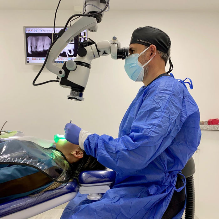
A properly configured microscope allows the dentist to maintain a more upright working position while viewing the operative field through the optical system.
The advantage isn't simply comfort during one procedure. Better positioning can support concentration and consistency across a full clinical schedule.
Dental Microscope vs Loupes
The dental microscope vs loupes discussion is often presented as a competition, but the two systems serve different working preferences and requirements.
Loupes offer portability and familiar magnification for a wide range of procedures. A microscope provides a broader range of magnification, more controlled illumination, and a stable viewing position.
For clinicians working routinely at higher magnifications or performing highly detailed restorative, cosmetic, or endodontic procedures, those differences can become significant.
The better question isn't necessarily which is better. It's where the additional visual and ergonomic control of a microscope justifies its place in the workflow.
How Documentation Helps Patient Communication
One of the less obvious advantages of modern dental microscopy is what happens beyond the operative field.
When a microscope is equipped with imaging capabilities, clinicians can capture photographs or video during treatment. That documentation can become useful for case review, clinical records, education, and patient conversations.
Instead of explaining a preparation margin or a structural concern verbally, the dentist can show the patient what they are seeing.
That changes the conversation.
Complex treatment becomes easier to understand when patients can see the condition being discussed and the work being performed.
In practices where patient communication plays a significant role in treatment planning, visual documentation can also contribute to case acceptance by making the value of recommended treatment more tangible.
Where Labomed Fits Into Modern Dental Microscopy
For more than six decades, Labomed has developed microscopy solutions for professionals who work where visibility and precision matter.
Its dental microscopy systems are designed around a combination of high-quality optics, illumination, ergonomics, and mechanical control.
The Labomed Magna, for example, provides continuous optical magnification from 4× to 25×, allowing clinicians to move through different levels of magnification without compromising the optical image. Its integrated LED illumination provides bright, consistent lighting, while the articulated design supports flexible positioning within the operatory.
The electromagnetic clutch system is designed to make positioning more intuitive, reducing the learning curve associated with microscope handling.
The result is a compact system built around a straightforward principle: the microscope should support the clinician's workflow rather than become another thing the clinician has to work around.
For restorative and cosmetic dentistry, that means bringing greater visual control into the procedures where small details matter most.
FAQs
Q. Is a dental microscope better than loupes for restorative dentistry?
A. It depends on the procedure and the clinician's workflow. Loupes provide portability and useful magnification, while a dental microscope offers higher and continuously adjustable magnification, integrated illumination, and a more ergonomic viewing position. For highly detailed restorative procedures, the additional control of a microscope can be particularly valuable.
Q. Do dental microscopes help with fillings and crowns?
A. Yes. Magnification can help clinicians evaluate preparation margins, tooth surfaces, restoration interfaces, and finishing details. This can support greater root canal precision, tooth preparation accuracy, and overall restorative control.
Q. Can a dental microscope help with veneers?
A. A microscope can support veneer procedures by improving visualization during tooth preparation, margin refinement, finishing, and evaluation of the restoration interface. It is particularly useful when small aesthetic and structural details need close inspection.
Q. Do dental microscopes reduce neck and back pain for dentists?
A. A microscope can support a more upright working posture by allowing the dentist to view the operative field through the optical system rather than leaning toward the patient to obtain a closer view. Proper operatory setup and positioning remain important to achieving the ergonomic benefit.
Q. Can a dental microscope help detect cracked teeth?
A. Higher magnification and focused illumination can make fine structural details easier to examine. However, a microscope is one diagnostic aid among several and does not independently determine whether a tooth is cracked.
Q. Can a dental microscope be used to show patients what the dentist sees?
A. Yes. When connected to a camera or imaging system, a microscope can display or record the operative field. This can support patient communication, case documentation, treatment discussions, and clinical education.
Q. What magnification level is used for dental procedures?
A. There is no single magnification appropriate for every procedure. Different stages of treatment call for different levels. A system with continuous magnification allows the clinician to move between lower magnification for orientation and higher magnification for detailed work.
Q. What is a dental microscope used for?
A. A dental microscope is used to improve visualization and control during precision procedures, including restorative dentistry, cosmetic dentistry, endodontics, tooth preparation, finishing, and other treatments where fine anatomical or procedural details matter.
Explore Labomed dental microscopes designed for high-precision visualization, ergonomic working, and greater control across restorative, cosmetic, and endodontic procedures.
Conclusion
In restorative and cosmetic dentistry, precision rarely comes down to one dramatic moment. It is built through dozens of small decisions made throughout a procedure.
The right level of magnification makes those decisions easier to evaluate. Better illumination brings greater clarity to the working field. Ergonomic design allows the clinician to maintain that precision without constantly adapting their posture.
That is ultimately where a dental microscope earns its place.
It doesn't replace experience or clinical judgment. It gives experienced clinicians a clearer view through which to apply both.
For practices focused on restorative quality, cosmetic detail, and minimally invasive treatment, microscopy can become a natural extension of the way they already work: with greater visibility, control, and precision.

© 2010-2026 Labo America, Inc. All Rights Reserved.